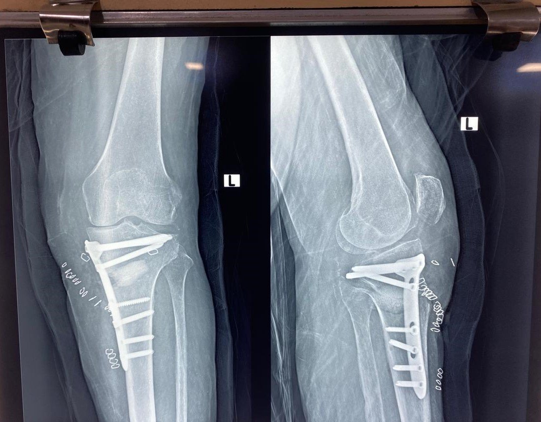

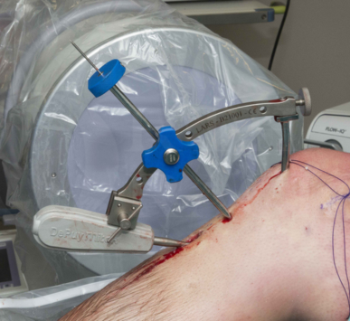
The cruciate ligaments are the central stabilizers of the human knee. The Anterior Cruciate Ligament (ACL) prevents the shin bone (tibia) from sliding excessively forward and controls rotational twisting, while the Posterior Cruciate Ligament (PCL) prevents the tibia from sagging backward. Injuries to these crucial ligaments are among the most common and disabling sports-related trauma, typically resulting from sudden decelerations, pivoting movements, or direct vehicular impacts.
Classic Symptoms of a Cruciate Ligament Tear
When an ACL tear occurs, patients typically report:
- An audible or felt 'pop' deep within the knee at the moment of injury.
- Rapid knee swelling (hemarthrosis) developing within 2 to 4 hours.
- Intense acute pain and immediate inability to continue playing or weight-bearing.
- Chronic sensation of knee instability—a feeling that the knee 'gives way' or buckles during pivoting or descending stairs.
Accurate Diagnosis & The Lachman Test
Dr. Emad Ansari conducts comprehensive clinical evaluations including the Lachman test, Anterior Drawer test, and Pivot Shift examination to assess rotational laxity. A high-resolution 1.5T or 3.0T MRI scan is performed to confirm the tear's grade (partial vs. complete) and evaluate associated injuries to the meniscus or articular cartilage.
Arthroscopic Anatomical Cruciate Reconstruction
Because the intra-articular synovial environment prevents torn cruciate ligaments from healing on their own, active patients with functional instability require arthroscopic reconstruction:
- Autograft Harvesting: A small, strong tendon—typically the hamstring (semitendinosus/gracilis) or bone-patellar tendon-bone—is harvested through a tiny incision.
- Tunnel Drilling: Under HD arthroscopic guidance, precise anatomical tunnels are drilled in the exact footprint where the original ligament attached.
- Graft Fixation: The prepared graft is passed through the tunnels and secured using high-strength titanium adjustable loop buttons and bioabsorbable interference screws.
Structured Rehabilitation & Return-to-Play Protocol
Successful ligament surgery is 50% surgical precision and 50% dedicated rehabilitation. Dr. Ansari prescribes a phased rehabilitation timeline: immediate range of motion exercises, closed-kinetic chain strengthening by month 2, running drills by month 4, and objective functional agility testing before clearing athletes to return to contact sports at 8 to 9 months.
Authored by Dr. Emad Ansari
MBBS, MS Ortho, FIJR — Consultant Orthopaedic, Joint Replacement & Trauma Surgeon, Bhiwandi, Maharashtra.
Providing evidence-based orthopedic care, minimally invasive trauma surgery, and comprehensive joint restoration across Maharashtra.